Science Highlights, October 31, 2018
Earth and Environmental Sciences
Researchers tackle the multifaceted impacts of increasing temperatures on forests
Educational Outreach
Team completes 45th Advanced Homemade Explosives Course, achieves Program of Record
Claire Sanders and Scott Cram lecture at the 41st Annual Course in Cytometry
Ricardo Lebensohn teaches Texas A&M’s Summer School on Computational Materials Science
Materials Physics and Applications
3D x-ray diffraction images dislocations in polycrystalline metals under tensile loading
Materials Science and Technology
Fusion-relevant simulations show new helium bubble growth mode in tungsten
Earth and Environmental Sciences
Researchers tackle the multifaceted impacts of increasing temperatures on forests
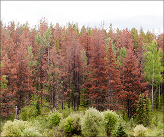
Photo. North American bark beetle outbreaks have killed millions of trees.
Forests cover one third of Earth’s total land area and contain more carbon in biomass and soils than is stored in the atmosphere. Every forested continent has undergone widespread, drought-induced forest mortality over the last two decades. Increasing temperatures could cause even greater stress on forests by superimposing longer periods of low precipitation on elevated evaporative demand (vapor pressure deficit). These environmental changes could impact populations of insect pathogens, increasing uncertainty regarding the fate of forests and projections of future carbon balance.
Lab researchers and collaborators tackled this problem through a multiscale model-experiment (ModEx) approach from cell to ecosystem to globe. A series of publications reporting their findings supports the Lab’s Energy Security mission area and Science of Signatures science pillar.
Universal assumptions about photosynthesis collapse under hot drought
Global carbon and water cycles, food production, and ecosystem services are dependent upon the diffusion of carbon dioxide (CO2) and water vapor through leaf pores. Current mathematical models of this process use the untested assumption that relative humidity (RH) inside leaves remains saturated under all conditions.
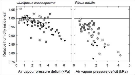
Figure. Relative humidity in leaf intercellular air spaces of juniper and piñon pine semiarid conifer species as a function of vapor pressure deficit. Relative humidity of one indicates saturation, denoted by horizontal line.
Experimentalists used a novel technique to couple measurements of leaf gas exchange and stable isotope compositions of CO2 and water vapor. In both mature conifer species measured, RH routinely dropped below saturation when leaves were exposed to increased vapor pressure deficits, which increase exponentially with increasing temperature. Correcting the bias caused by assuming saturated RH under all conditions will enable improved prediction of plant function and contribution to global carbon and water cycles under warmer temperatures.
Reference: “Unsaturation of Vapor Pressure Inside Leaves of Two Conifer Species,” Scientific Reports 8, 7667 (2018); DOI: 10.1038/s41598-018-25838-2. Authors: Lucas A. Cernusak (James Cook University), Nerea Ubierna, Suan Chin Wong, and Graham Farquhar (Australian National University); Michael Jenkins (University of California – Santa Cruz); Steven Garrity (METER Group); Thom Rahn, Heath Powers, and Sanna Sevanto (Earth System Observations, EES-14); David Hanson (University of New Mexico); and Nate McDowell (Pacific Northwest National Laboratory). The DOE Office of Science, Biology and Environmental Research (BER) program funded the work at Los Alamos. Technical contact: Sanna Sevanto
First 3D plant tissue imaging reveals important links between structure and drought response
Plants take up CO2 from the atmosphere for photosynthesis and control simultaneous water loss with the stomata (leaf pores). Plants close stomata during drought to avoid excessive water loss, which could lead to embolism and catastrophic failure of the water conducting system.
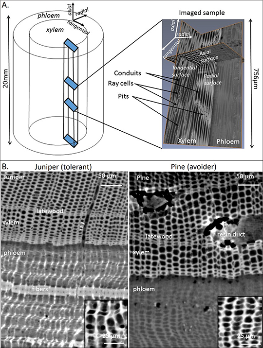
Figure. A) Schematic of samples imaged with X-ray tomography and orientation of different planes with example of 3D rendered image (right). B) Axial views of one-seed juniper (left) and piñon pine (right) tissues at the xylem-phloem transition.
The drought severity at which stomata close is related to structure of the woody water-transport tissue (xylem) and its vulnerability to embolism. It may also be related to structure of the soft sugar-transport tissue (phloem), allowing for sugar translocation under varying degrees of drought. Researchers conducted the world’s first experiment using tridimensional synchrotron X-ray microtomography and light microscopy of xylem and phloem tissues to evaluate changes in anatomy of two coniferous species with contrasting xylem vulnerabilities, desiccation tolerance, and stomatal closure points. The unique images reveal that desiccation tolerant plants require higher phloem transport capacity than desiccation avoiding plants.
Reference: “Is Desiccation Tolerance and Avoidance Reflected in Xylem and Phloem Anatomy of Two Co-Existing Arid-Zone Coniferous Trees?” Plant, Cell & Environment 41, 1551 (2018); https://doi.org/10.1111/pce.13198. Authors: Sanna Sevanto, Max Ryan, and L. Turin Dickman (EES-14); Dominique Derome (Empa, Switzerland); Alessandra Patera (Paul Scherrer Institute, Switzerland); Thijs Defraeye (Empa; ETH Zurich, Switzerland); Robert Pangle, Patrick Hudson, and William Pockman (University of New Mexico). The DOE BER program funded the experiment from which the samples were collected, and Laboratory Directed Research and Development (LDRD) funded the imaging and analysis at Los Alamos. Technical contact: Sanna Sevanto
Computational method adds stochastic insect and plant phenology in environment simulations
Warming temperatures alter the phenology (periodic plant and animal life cycle events) of plants and insects. Many plants flower sooner and many insects emerge earlier than they did in the previous decade. Accurate representation and prediction of these changes requires a stochastic representation to capture phenotypic and environmental variability across regions and through time.
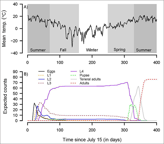
Figure. A) Model predictions for mountain pine beetle development (phenology) forced using observed seasonal temperatures recorded in Jasper National Park, Alberta, Canada. B) Expected number of individuals in each of the stages as determined by the stage- and age-structured model.
Researchers have developed a method to capture the effects of stochasticity without requiring ensembles of stochastic simulations, which can be computationally expensive for global earth system models. The team demonstrated the approach using a temperature-dependent model of the demography of the mountain pine beetle, an insect that kills mature pine trees. Many of the temperature-based phenology models traditionally used by scientists could be efficiently incorporated in earth system models using this approach.
Reference: “Incorporating Variability in Simulations of Seasonally Forced Phenology using Integral Projection Models,” Ecology and Evolution 8, 162 (2018); DOI: 10.1002/ece3.3590. Authors: Devin Goodsman (EES-14, University of Alberta), Brian Aukema (University of Minnesota), Nate McDowell (Pacific Northwest National Laboratory), Richard Middleton (Computational Earth Science, EES-16), and Chonggang Xu (EES-14). The Laboratory Center for Earth and Space Science funded the model development, and the LDRD program sponsored the data analysis at Los Alamos. Technical contact: Chonggang Xu
Impact of forest canopy architecture on ecosystem carbon, water, and energy exchange
Large-scale earth system models only consider a single-layer view of the forest canopy. However, the vertical and horizontal structure of the canopy may also be important for accurately capturing ecosystem fluxes. Researchers used a leaf level plant biophysics model that solves photosynthesis, stomatal control, leaf-atmosphere gas exchange, and energy balance equations with analytical solutions of wind flow and light attenuation inside the canopy to explore effects of canopy architecture on these parameters. For the same forest canopy coverage fraction or leaf area index, they found that different vertical organizations of foliage density lead to different microclimates inside the canopy by modulating wind turbulence, light and other meteorological parameters. This leads to significant differences in ecosystem level fluxes of heat, water (transpiration), and carbon. These aggregate effects are essential for accurately capturing earth system dynamics. This information could have potential applications in agriculture and sustainability practices for healthy forests.
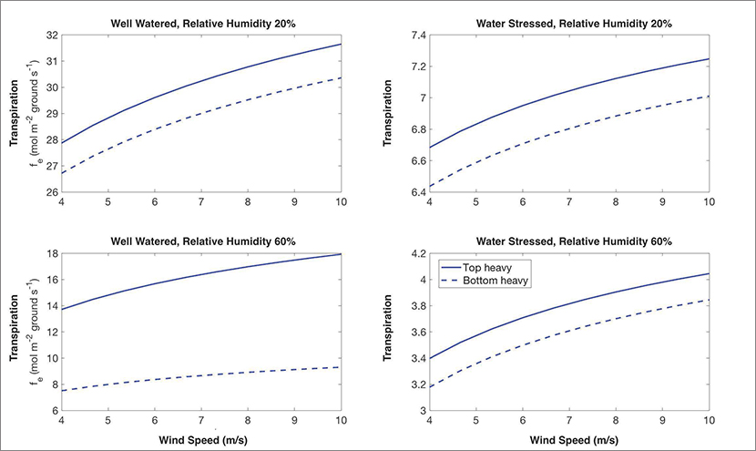
Figure. Variation of net canopy transpiration with tree-top wind speed for two theoretical species under four different conditions of soil and atmospheric water availability. The two theoretical tree species differ in vertical leaf distribution, but have the same amount of total foliage. Therefore, a large-scale earth system model would not differentiate between them.
Reference: “Effect of Vertical Canopy Architecture on Transpiration, Thermoregulation and Carbon Assimilation,” Forests 9, 198 (2018); DOI: 10.3390/f9040198. Authors: Tirtha Banerjee and Rodman Linn (EES-16). A Laboratory Center for Earth and Space Science Chick Keller Postdoc Fellowship and a Director’s Fellowship funded the research. Technical contact: Tirtha Banerjee
Competition for trees counteracts effect of warmer temperatures on bark beetle populations
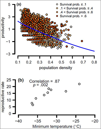
Figure. Contrasting effects of population density and temperature on bark beetle populations. Bark beetle productivity declines with population density at the site level (a), counteracting the positive effects of warmer winter temperatures on bark beetle reproductive rates (b).
Bark beetles can cause extensive forest mortality, and warmer temperatures could increase bark beetle outbreak frequency, severity, and range. However, resource limitation could cause outbreaks to decelerate. Researchers have used an extensive 9-year dataset, in which nearly 10,000 trees in Alberta were sampled across a region of approximately 90,000 km2 recently invaded by the mountain pine beetle, to evaluate how temperature could impact mountain pine beetle reproduction. Global Change Biology reported their findings.
Their analysis supports a positive effect of warmer winter temperatures on mountain pine beetle overwinter survival at the landscape and regional scales (top Figure panel). However, this effect is overwhelmed by competition for food resources under tree bark at the site level (bottom Figure panel). The team’s research suggests that accounting for negative density dependence (overcrowding) due to competition for resources is necessary to determine the effects of warming temperatures on bark beetle populations at small spatial scales. The authors conclude that sustained bark beetle outbreaks in warming temperature regimes will depend on the amount of their dispersal from overcrowded regions.
Reference: “The Effect of Warmer Winters on the Demography of an Outbreak Insect Is Hidden by Intraspecific Competition,” Global Change Biology 24, 3620 (2018); DOI: 10.1111/gcb.14284. Authors: Devin Goodsman (EES-14 and University of Alberta), Guenchik Grosklos (Utah State University), Brian Aukema (University of Minnesota), Caroline Whitehouse (Alberta Agriculture and Forestry), Katherine Bleiker (Canadian Forest Service), Nate McDowell (Pacific Northwest National Laboratory), Richard Middleton (EES-16), and Chonggang Xu (EES-14). The Lab Center for Earth and Space Science funded model development, and LDRD sponsored data analysis at Los Alamos. Technical contact: Chonggang Xu
Educational Outreach
Team completes 45th Advanced Homemade Explosives Course, achieves Program of Record
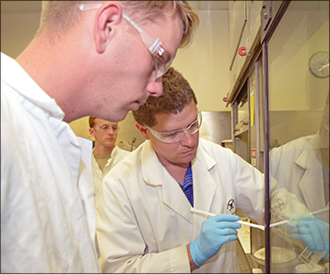
Photo. Bryce Tappan teaches the primaries synthesis laboratory to students behind a blast shield.

Photo. Students compare witness plates following performance testing of materials prepared in the class.
On October 19, a diverse team of scientists, technologists, technicians, and professional staff completed the 45th offering of the LANL Advanced Homemade Explosives (HME) Course, which has been awarded Program of Record funding through the Navy Center for EOD (Explosive Ordnance Disposal) and Diving. The primary objective of the class is to provide EOD technicians with the advanced knowledge and critical thinking necessary to identify and handle HMEs safely. Five days of hands-on training at the Lab emphasize safety and awareness through HME synthesis and formulation, HME performance, and HME detection. The team has trained more than 800 students and is scheduled to complete 9 classes in both 2018 and 2019, training as many as 24 EOD technicians per class.
The Lab developed the hands-on training course in 2012 after the Department of Defense expressed a need for safe training for EOD technicians. The Laboratory team was nearing completion of a series of three-day courses in HME situational awareness for the Pentagon’s Joint Improvised Explosive Device Defeat Organization (JIEDDO). The JIEDDO provided the initial funding for the development and first few years of training for what is now the five-day hands-on LANL Advanced HME Explosives Course.
Since its inception during the Manhattan Project, the Laboratory has developed cradle-to-grave expertise in explosives development, characterization, and testing. The Advanced HME Class Team leverages 75 years of expertise in comprehensive explosives development. Co-principal investigators Virginia Manner (HE Science and Technology, M-7), Margo Greenfield (Shock and Detonation Physics, M-9), and Jackie Veauthier (Inorganic, Isotope and Actinide Chemistry, C-IIAC) have led the HME course. Current team members include J. AlderseBaes and J. Whitton (Intelligence and Systems Analysis, A-2), J. Veauthier (C-IIAC), D. Oschwald (Physical Chemistry and Applied Spectroscopy, C-PCS), A. Cartelli, C. Campbell, M. Campbell, and A. Novak (Explosives Applications and Special Projects, M-6); S. Anthony, P. Bowden, G. Brown, D. Chavez, A. Duque, L. Fleming, E. Hartline, P. Leonard, V. Manner, D. McDonald, A. Schmalzer, C. Snyder, B. Tappan, and K. Windler (M-7); K. Brown, M. Greenfield, S. McGrane, D. Moore, and K. Paul (M-9); M. Quartieri (Explosive Science and Shock Physics, M-DO); W. Boncher (W88 Alteration and Refresh Program, Q-17); and C. Johansen (Emergency Response, EO-ER). The National Security and Defense Program Office manages the course.
The course has received several awards and recognition through the Los Alamos Award Program, the NNSA Defense Programs Awards of Excellence, and in two LANL press release videos (https://www.lanl.gov/projects/explosives-detection/capabilities/training.php). The DoD Center for Explosives Ordnance Disposal and Diving funds the class. The work supports the Lab’s Global Security mission area and the Science of Signatures and Materials for the Future science pillars. Technical contacts: Virginia Manner and Jackie Veauthier
Claire Sanders and Scott Cram lecture at the 41st Annual Course in Cytometry

Photo. Claire Sanders teaching at the 41st annual Flow Course.
Los Alamos developed the flow cytometry cell separation technique in the 1960s. The Lab has continued to advance technology development as well as promote education and training – primarily through its role in the NIH-funded National Flow Cytometry Sorting and Research Resource that was housed at Los Alamos from 1983-2014. One of the main elements of the training program has been the Annual Course in Cytometry, which is now in its 41st year. Clair Sanders (B-11) and Lab retiree Scott Cram taught sections of this year’s course in Bowdoin, ME.
Sanders has taught laboratory sessions at eight courses with Cram, who has received the Los Alamos Medal for his pioneering use of flow cytometry to isolate chromosomes for the Human Genome Project. In their hands-on lab Sanders and Cram walked the students through the isolation, staining, and flow cytometric analysis of mammalian chromosomes from hamster cells in culture. Students learned the specifics of this procedure, instrument optimization, and small particles analysis.
The work supports the Lab’s Global Security mission area and the Science of Signatures science pillar. Technical contact: Claire Sanders
Ricardo Lebensohn teaches Texas A&M’s Summer School on Computational Materials Science
Photo. Ricardo Lebensohn lecturing.
The Texas A&M University Computational Materials Science Across Scales program provides knowledge exchange for academics and trains graduate students. The annual summer school offers a thorough overview of some of the most important computational tools currently in use to investigate materials phenomena at multiple scales, ranging from the continuum to the electronic structure level. The course also presents the latest advances in materials informatics for materials discovery and design. The two-week long summer school took place at Texas A&M in College Station, TX.
Ricardo Lebensohn (Fluid Dynamics and Solid Mechanics, T-3) taught the school’s “Polycrystal Plasticity” module. He focused on the theories and corresponding numerical codes developed at the Lab for physically-based modelling of the mechanical behavior of polycrystalline aggregates. Polycrystals are a class of materials whose complexity requires the use of models at different spatial and temporal scales for a quantitatively-accurate and microstructure-sensitive prediction of their mechanical properties. Lebensohn, along with a group of distinguished scientists and scholars, is also member the summer school’s advisory board.
Lebensohn has worked in the area of structure/property relationship of polycrystalline materials for more than 25 years and is an expert in crystal plasticity modelling. His contributions include the viscoplastic self-consistent (VPSC) code, a homogenization-based polycrystal plasticity simulation tool for the prediction of mechanical response and microstructure evolution of polycrystalline aggregates and Fast Fourier Transform (FFT)-based codes, for the prediction of micromechanical fields in experimental mechanics. His work supports the Lab’s Energy Security Mission area and the Materials for the Future science pillar. Technical contact: Ricardo Lebensohn
Materials Physics and Applications
3D x-ray diffraction images dislocations in polycrystalline metals under tensile loading
The nucleation and propagation of dislocations is a ubiquitous process that accompanies the plastic deformation of materials. Material defects and the strain fields accompanying them influence a range of material’s properties, e.g., from mechanical strength to failure, battery performance, radiation resistance, and crystal growth and dissolution. Researchers have sought to tailor material response through defect engineering and control since these defects were first visualized more than 50 years ago using transmission electron microscopy. A team from Los Alamos and Argonne national laboratories have demonstrated a novel advanced imaging technique that offers the promise of accomplishing this more effectively by applying nanometer-scale x-ray strain imaging to materials being pulled apart. Nature Communications published their findings.
The team used Bragg coherent x-ray diffraction imaging (BCDI) at Argonne’s Advanced Photon Source (APS) to measure the strain field of a free-standing polycrystalline film after tensile loading. The researchers integrated the observed three-dimensional structure into atomistic modeling to show that the measured strain field corresponds to a screw dislocation – a specific form of a dislocation in the crystal’s lattice structure. This is the first example of direct 3D x-ray imaging of the strain field surround a line defect within a grain of unsupported nanocrystalline material following tensile loading. Moreover, this is the first demonstration of a framework that enables complementary atomistic simulations to be performed on structure experimentally imaged through Bragg coherent x-ray diffraction measurements. This integration of experiments and atomistic modeling could be leveraged to provide insight into dynamic processes that each method alone cannot accomplish.
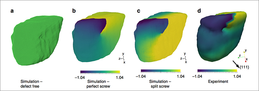
Figure. Atomic structure and displacement fields with and without a screw dislocation. a) Atomistic structure obtained by filling copper atoms into the experimentally observed grain. b) Atomistic structure following addition of a perfect screw dislocation, and c) atomistic structure after energy minimization. Atoms are colored by their displacement along the (111) crystallographic direction from their initial structure. c) isosurface rendering of the experimentally imaged copper grain.
The work supports the Laboratory’s Nuclear Deterrence and Global Security mission areas and its Materials for the Future science pillar, by advancing efforts to develop materials with controlled functional and predictable performance, a key objective of the Lab’s materials strategy. The ability to visualize locations and long-range strain fields is vital to the improved design of functional and structural materials with pre-engineered defects. The research shows the insight enabled by the use of high-energy, advanced light sources, such as the APS and the Lab’s proposed MaRIE capability, to probe the mesoscale – the spatial scale where a material’s structure strongly influences its macroscopic behaviors and properties. MaRIE is the Laboratory’s proposed facility to study matter-radiation interactions in extremes. This work also falls under a partner user proposal with researchers Ross Harder and team at the Advanced Photon Source where the joint Los Alamos APS teams are applying these techniques to study the response of bulk materials to damage.
Reference: “Three-dimensional X-ray Diffraction Imaging of Dislocations in Polycrystalline Metals under Tensile Loading,” Nature Communications 9, 3776 (2018); DOI: 10.1038/s41467-018-06166-5. Authors: Mathew J. Cherukara, Evan Maxey, Wonsuk Cha, Jorg Maser, and Ross J. Harder (Argonne National Laboratory); Reeju Pokharel and Saryu J. Fensin (Materials Science and Technology, MST-8); Timothy S. O’Leary and J. Kevin Baldwin and Richard L. Sandberg (Center for Integrated Nanotechnologies, MPA-CINT).
The Laboratory Directed Research and Development (LDRD) program, the Dynamic Materials Properties Campaign (C2, Lab Program Manager Dana Dattelbaum) of the Laboratory’s Experimental Sciences Program, and the Center for Integrated Nanotechnologies, a DOE Office of Basic Energy Sciences user facility jointly operated by Sandia National Laboratories and Los Alamos National Laboratory sponsored different aspects of the work at Los Alamos. Technical contact: Richard Sandberg
Materials Science and Technology
Fusion-relevant simulations show new helium bubble growth mode in tungsten
The design of materials that withstand the extreme radiative environment in fusion reactors presents a significant technical outstanding challenge. Due to its excellent material properties, tungsten (W) is a leading material choice for plasma-facing material in these reactors. As a reactor component, tungsten will be implanted with a high flux of low energy helium ions. However, helium implantation causes morphological changes that might disrupt the material’s performance and lead to contamination of the plasma. A team from Los Alamos and Oak Ridge National Laboratory (ORNL) used simulations of fusion-relevant conditions to reveal a new helium bubble growth mode in tungsten. The journal Materials Research Letters published their findings.
The study relied on ORNL’s Titan high-performance computing clusters, which provided about 5,000 CPUs for a total of about two million CPU hours. The researchers created models of accelerated molecular dynamics of helium bubble nucleation and growth at tungsten Σ5(310)[001] grain boundaries. In the simulation, helium interstitial atoms (or helium atoms that exist in a gap between crystal lattice sites) migrated slowly at grain boundaries (about 1–3 ns per hop) and then nucleated bubbles by punching of interstitials. The figure shows the simulations for helium implantation at a rate of 108 helium atoms per second (left panel) and a rate of 107 helium atoms per second (right panel). Thermodynamic interactions with the bubble itself prevented the interstitials from escaping. This caused the interstitial halo to block helium supply to the bubble.
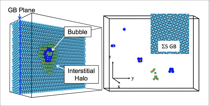
Figure. (Left): Helium bubble growth at a tungsten grain boundary (GB). The halo of tungsten interstitials around the helium bubble constrains helium arrival and thus bubble growth, with a limited maximum size. (Right): A snapshot of bubble nucleation at the grain boundary shows nucleation of helium clusters containing 6, 4, and 2 helium atoms (blue spheres). Green and orange spheres indicate defects, and cyan spheres are the original tungsten atoms.
The team showed that this novel growth mode — which involved a halo forming around the growing bubble — reduced the bubble’s helium supply and therefore arrested bubble growth. The researchers concluded that the kinetics associated with bubble growth at grain boundaries in tungsten is qualitatively different from that in the bulk. Understanding how helium bubbles nucleate and grow is important for predicting the large-scale evolution of the material, which could help commercialize fusion power by developing plasma-facing materials that tolerate the extreme conditions of elevated temperatures and high particle flux in fusion reactors. This research advances the knowledge of helium evolution at grain boundaries in tungsten, an important consideration for understanding tungsten as a plasma-facing component in fusion reactors.
Reference: “New Helium Bubble Growth Mode at a Symmetric Grain-Boundary in Tungsten: Accelerated Molecular Dynamics Study,” Materials Research Letters 6,:9, 522 (2018); DOI: 10.1080/21663831.2018.1494637. Authors: X.-Y. Liu and B. P. Uberuaga (Materials Science in Radiation and Dynamics Extremes, MST-8), D. Perez and A. F. Voter (Physics and Chemistry of Materials, T-1).
The DOE Office of Fusion Energy Sciences, Office of Basic Energy Sciences’ Materials Sciences and Engineering Division, and Office of Advanced Scientific Computing Research through the Scientific Discovery through Advanced Computing (SciDAC-4) program funded the research at Los Alamos. The team performed research at Los Alamos and computing at ORNL. The work supports the Laboratory’s Energy Security science mission area and its Materials for the Future and Information, Science, and Technology science pillars through the development of materials for fusion reactors. Technical contact: Xiang-Yang (Ben) Liu
Physics
Neutron lifetime measured with unprecedented precision
A free neutron decays with a lifetime of about 15 minutes to form a proton, electron, and antineutrino. Knowledge of the mean neutron lifetime (τn) helps scientists predict the atomic makeup of the early universe and search for physics beyond the Standard Model of particle physics. Measuring the exact lifetime of neutrons is surprisingly difficult. The τn measurements vary depending on the method used, and the potential corrections have always been larger than the uncertainties. The journal Science has published findings from researchers at Los Alamos and their collaborators of the first modern τn measurement with corrections that are smaller than the range of uncertainty. The study paves the way for better understanding of how atoms were first created during the Big Bang.
Researchers at the Los Alamos Neutron Science Center (LANSCE) developed the diagnostics – including the asymmetric trap and the detector – and new techniques required to perform this experiment. The team used the 800-MeV proton beam and ultracold neutrons produced at the Ultracold Neutron Facility at LANSCE to develop an improved method for measuring τn. The ultracold neutrons are of such low energies that they travel only a few meters per second. For this “trap” method, researchers levitated ultracold polarized neutrons in a container using magnetic fields and gravity, holding them in place for as long as an hour until a detector was placed in situ. The detector counted neutrons that had not decayed by determining which particles were reabsorbed into matter. There was no detectable neutron loss caused by interactions with the magnetic and gravitational walls of the trap.
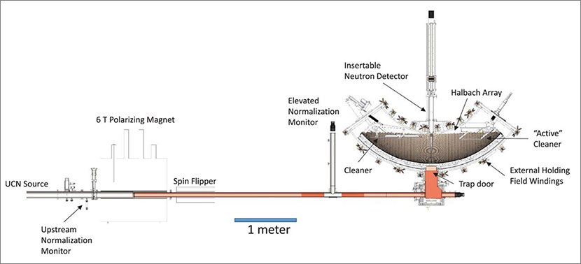
Figure. Layout of the ultracold neutron beam line and trap used for these measurements.
The results of previous measurement techniques have historically disagreed by 9.2 seconds. The new method has narrowed the systematic uncertainty to +0.4/–0.2 seconds. This experiment marks the first time that neutron lifetime measurements did not require corrections larger than the quoted uncertainties. It is also the first time that researchers have used an in situ neutron detector to count neutrons in a trap. Because the study’s measured uncertainties appear to be statistically driven, the team predicts that future studies using this apparatus could reduce the uncertainty of the mean neutron lifetime to well below 0.5 seconds.
Reference: “Measurement of the Neutron Lifetime using a Magneto-gravitational Trap and in situ Detection,” Science 360, 627 (2018); DOI: 10.1126/science.aan8895. Authors: R. W. Pattie Jr., C. Cude-Woods, S. M. Clayton, S. A. Currie, D. E. Fellers, S. W. T. MacDonald, M. Makela, C. L. Morris, J. D. Ortiz, J. Ramsey, A. Saunders, Z. Tang, Z. Wang, W. Wei, H. L. Weaver, and B. A. Zeck (Subatomic Physics (P-25); M. A. Hoffbauer (Chemical Diagnostics and Engineering, C-CDE); S. K. Sjue (Applied Modern Physics, P-21); P. L. Walstrom (Accelerators and Electrodynamics, AOT-AE); T. L. Womack (formerly P-25, now retired); and S. J. Seestrom (formerly at Los Alamos, currently at Sandia National Laboratories). External team members: E. R. Adamek, N. B. Callahan, W. Fox, C.-Y. Liu, and J. Vanderwerp (Indiana University); E. R. Dees, J. W. Wexler, and A. R. Young (Triangle Universities Nuclear Laboratory and North Carolina State University); L. J. Broussard (Oak Ridge National Laboratory); X. Ding (Virginia Polytechnic Institute and State University); E. M. Engel (West Point Military Academy); P. Geltenbort (Institute Laue-Langevin, Grenoble); K. P. Hickerson (California Institute of Technology); A. T. Holley (Tennessee Technological University); A. Komives (DePauw University); D. J. Salvat (University of Washington); and E. L. Sharapov (Joint Institute for Nuclear Research, Russia).
The Los Alamos Laboratory Directed Research and Development (LDRD) program funded the development of the concept and a prototype apparatus at Los Alamos, and the DOE Office of Science Office of Low Energy Nuclear Physics funded the experiments. The work supports the Laboratory’s Nuclear and Particle Futures science pillar by developing capabilities to measure the mean lifetime of the neutron. Technical contact: Andy Saunders
Sigma
Neutron diffraction reveals texture changes in low-enriched metallic uranium nuclear fuel
The United States is committed to further strengthening nuclear security and nonproliferation to reduce the threat of terrorists acquiring nuclear material. To meet this important mission, Los Alamos brings its expertise to the task of converting research reactors worldwide to use low-enriched uranium (LEU) fuels, which cannot be used in weapons, rather than highly enriched uranium (HEU) fuels. This work is part of the NNSA’s Office of Material Management and Minimization Reactor Conversion (CONVERT) program.
To aid the development of a widely applicable LEU fuel, Laboratory materials scientists examined the microstructural changes that occur during the fuel fabrication process, a relatively unexplored area of study. Their work revealed texture changes during processing – changes which affect the material’s properties. Understanding these properties is critical for efficiently and economically manufacturing LEU fuel. The journal Quantum Beam Science published their findings.
The team used high-density U-10 wt% Mo (a uranium-molybdenum alloy clad in 6061 aluminum) for their work. Researchers employed a Sigma Division-developed plasma-spray technique as a fast and economical coating method to apply a zirconium (Zr) coating that acts as a diffusion barrier between the fuel and aluminum cladding.
The investigators performed time-of-flight (ToF) neutron diffraction on the high-pressure-preferred orientation (HIPPO) instrument at the Los Alamos Neutron Science Center (LANSCE) to examine the microstructure evolution caused by various plasma temperatures. Neutrons are an excellent probe for bulk texture measurements of uranium and its alloys because they probe the entire sample volume. X-ray or electron-based methods only probe a few micrometers in these materials. The neutron diffraction method collects the bulk information of both substrate (U-10Mo, ~0.3 mm thickness) and thin coating (Zr, ~30 µm thickness) at the same time in a non-destructive way. The neutron diffraction data from HIPPO provides a wide variety of material information including crystallographic textures, phase fractions, lattice parameters, and microstrain.
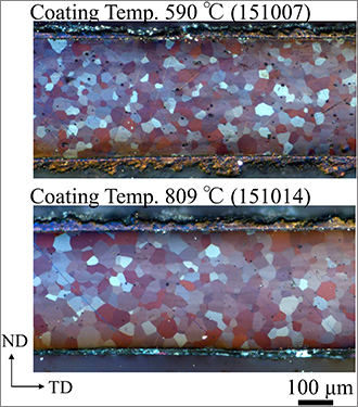
Figure. Optical micrographs of two Zr-coated samples: 151007 and 151014 coated at the lowest and highest temperature, respectively. Both samples showed equiaxed grains, suggesting a recrystallized microstructure, and the grains were coarser near the coating surface than in the center. ND is sample normal and TD is transverse direction.
Quantitative texture analysis revealed that the texture drastically changed at high coating temperatures. The team attributed this result to selective grain growth. This finding agrees with optical microscopy data. Researchers observed larger grains in the samples closer to the coated surface than in the unaffected center volume. For the surface regions exposed to plasma spraying, the average grain size for the lowest temperature agreed with the grain size at the center line. However, the grain size for the sample at the maximum temperature showed an increase of 27.5% relative to the center line grain size. The Zr coating indicated a preferential orientation, which could be related to the initial texture of the uncoated U-10Mo. The authors suggest that this orientation could be explained by epitaxial growth of Zr on the U-10Mo substrate.
Reference: “Texture Evolution in U-10Mo Nuclear Fuel Foils during Plasma Spray Coating with Zr,” Quantum Beam Science 2, 12 (2018); doi.org/10.3390/qubs2020012 Authors: Shigehiro Takajo and Sven C. Vogel (Materials Science in Radiation and Dynamics Extremes, MST-8), Kendall J. Hollis, Dustin R. Cummins and David E. Dombrowski (Fabrication Manufacturing Science, Sigma-1), and Eric L. Tegtmeier (Finishing Manufacturing Science, Sigma-2).
NNSA’s Office of Material Management and Minimization Reactor Conversion Program (Lab Program Manager Dave Dombrowski) funded the work, which supports the Laboratory’s Global and Energy Security mission areas and the Materials for the Future science pillar through the development of methods to produce LEU for reactors. The plasma spray technique leverages the manufacturing science expertise and capabilities of the Laboratory’s Sigma Complex, which supports a large, multidisciplinary materials technology base. The ToF diffraction studies employed capabilities at LANSCE, which uses intense pulsed protons to produce the wide energy spectrum of spallation neutrons needed to interrogate materials. Technical contact: Sven Vogel
Theoretical
A new approach to analyze cell signaling models demonstrated
Models in systems biology seek to describe how the concentrations of key signaling proteins within a cell change in response to a stimulus. Such models are important in understanding biological processes at the single-cell level, as well as predicting cellular responses to therapeutic treatment. To make accurate predictions, models require an acceptable choice of parameters – numerical quantities such as the concentration of proteins and rates of biochemical reactions. In a publication in Nature Communications, Eshan Mitra and Bill Hlavacek (Theoretical Biology and Biophysics, T-6) and collaborators from Northern Arizona University demonstrated an approach that improves cell signaling models by allowing new kinds of experimental data to inform the model parameters.
In a conventional approach, model parameters are fit to quantitative experimental data, such as a dose-response curve for a particular drug. The study demonstrated that it is also possible to include non-numerical, qualitative data in model fitting. The team showed that with a sufficiently large number of qualitative measurements, it is possible to improve quantitative confidence in model parameters. This finding is important in the field of cell biology, where quantitative experiments are often technically challenging to perform, and a large proportion of the available data in the literature is qualitative.
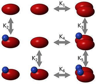
Figure. A simple model of dimerization and inhibition of a protein. Red ovals represent the protein Raf, and blue spheres represent an inhibitor of Raf. This type of model can benefit from qualitative experimental data by applying the methodology of the study.
The team demonstrated the new approach by fitting a model of the cell cycle in yeast. Yeast is used as a model organism to help understand the cell cycle in human cells, which is relevant to cancer. The experimental literature on yeast contains both quantitative data (RNA levels at different time points) and qualitative data (mutant yeast strains with known behaviors). The team implemented an optimization algorithm that found model parameters that were consistent with both types of data. The findings revealed that the combination of both data sets yielded a higher confidence in parameter values than either of the individual data sets.
The results create exciting prospects for future collaborations between experimental and theoretical biologists. By allowing theorists to utilize experimental data that are more readily generated, the new approach is a step toward more accurate, experimentally validated systems biology models.
Reference: “Using Both Qualitative and Quantitative Data in Parameter Identification for Systems Biology Models,” Nature Communications 9, 3901 (2018) DOI: 10.1038/s41467-018-06439-z. Authors: Eshan D. Mitra and William S. Hlavacek (Theoretical Biology and Biophysics, T-6), Raquel Dias Richard G. Posner (Northern Arizona University).
The National Institute of General Medical Sciences of the National Institutes of Health (NIH) funded Bill Hlavacek’s research, and the Laboratory Directed Research and Development (LDRD) program sponsored Eshan Mitra’s work. The research supports the Lab’s Global Security mission area and the Information, Science and Technology science pillar for applications in bio-security and public health. Technical contacts: Eshan Mitra and Bill Hlavacek
